Axis Medical Canada
West / Ouest: 1 888 855-6558 Ontario: 1 800 267-5597 East / Est: 1 877 388-1515
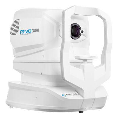
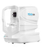
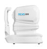
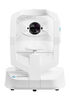
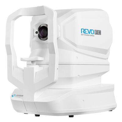
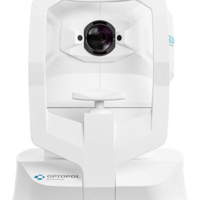
We proudly announce the REVO FC with AccuTrack - Real-Time hardware eye-tracking – the all-in-one OCT and a Fundus Camera. The device offers ultimate versatility for time and space efficiency, featuring full-fledged, high-resolution OCT with hardware eye-tracking and true color fundus imaging.
OPTOPOL pioneered the first Spectral Domain OCT technology 25 years ago. Their background allows them to provide an unparalleled experience to both patients and eye care professionals. Get the best of both worlds with ultra-high-quality scans in an easy-to-use interface.
AccuTrack
Hardware-based eye tracker compensates for blinks, loss of fixation, and involuntary eye movements during scans reducing artifacts.
Auto Functions
Simplifying operation with the push of a button to auto-position, auto-align, auto-focus, and auto-capture.
A.I. DeNoise
An advanced artificial intelligence (AI) algorithm removes noise from the tomogram for the highest image quality.
Custom Scan Protocols
Save time and never miss a scan. Create a custom preset group of scans and let the REVO capture all scans in order.
Motion Correction
The software-based motion correction (MC) compensates for involuntary eye movements and blinks by capturing two scans and generating a motion-corrected scan when necessary.
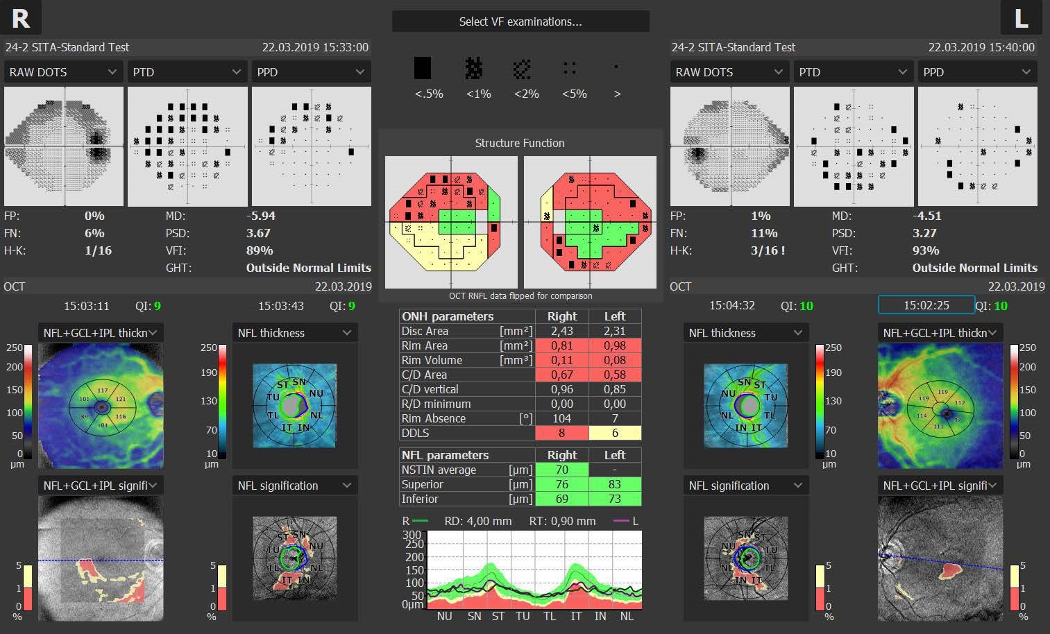
Structure + Function (S+F)
Comprehensive glaucoma solution that combines REVO OCT and PTS Visual Field results. S+F takes the diagnostic approach of the Hood report.
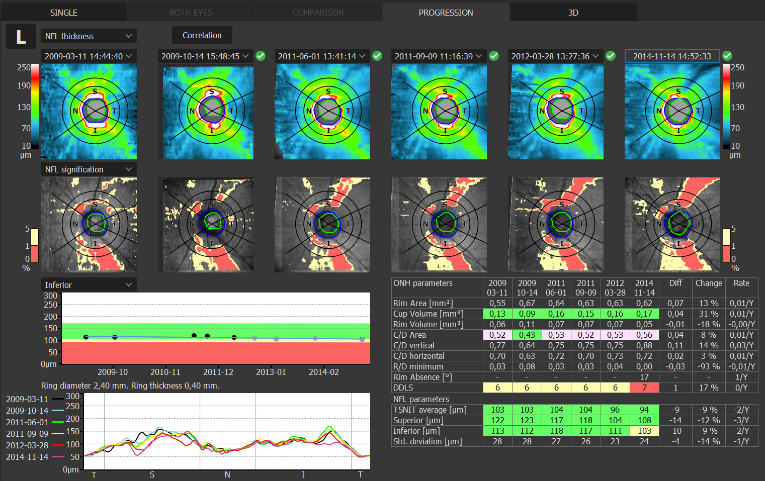
Progression Analysis
Gather baselines and follow-ups to monitor and manage disease progression in posterior and anterior scans.
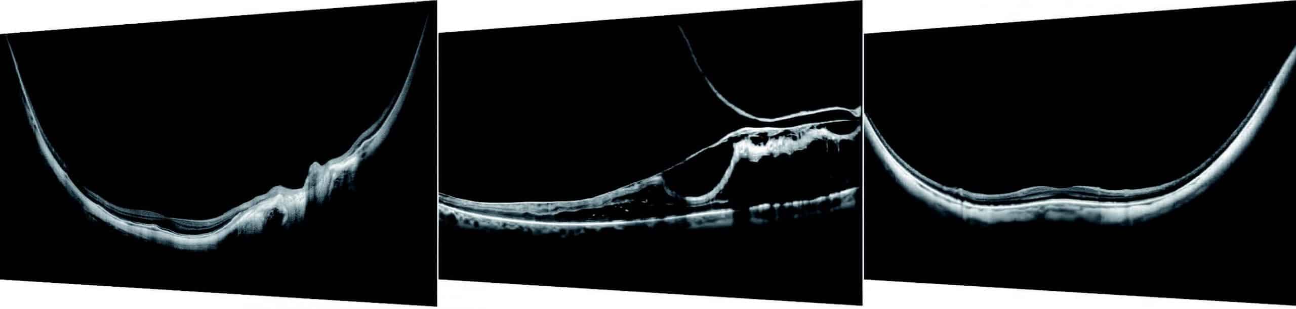
Full Range
With scans presenting New Extended DepthTM software, based on the Full Range technology, provides scans of increased depth for reliable and convenient observation of challenging cases. With scans presenting extended depth, this new imaging mode is perfect for diagnosing even highly myopic patients.
Progression Analysis
Quickly view a chronological set of exams for analysis of changes in morphology, quantified progression maps, and progression trends.
Custom Scan Protocols
Save time and never miss a scan. Combine any scan type into a pre-set group. Choose a group of scans and set the order, the REVO will do the rest.
Glaucoma Tool Kit
Comprehensive glaucoma analytical tools for quantification of the Nerve Fiber Layer and Ganglion Cell Layer. The Disc Damage Likelihood Scale enables clinicians to precisely diagnose and monitor glaucoma for Optic Nerve Head analysis.
Structure + Function
The S+F report allows clinicians to understand the relationship between structural glaucoma damage and the functional impact on the patient’s field of vision. This provides a quick and comprehensive single-page report for glaucoma management.
(sold separately)
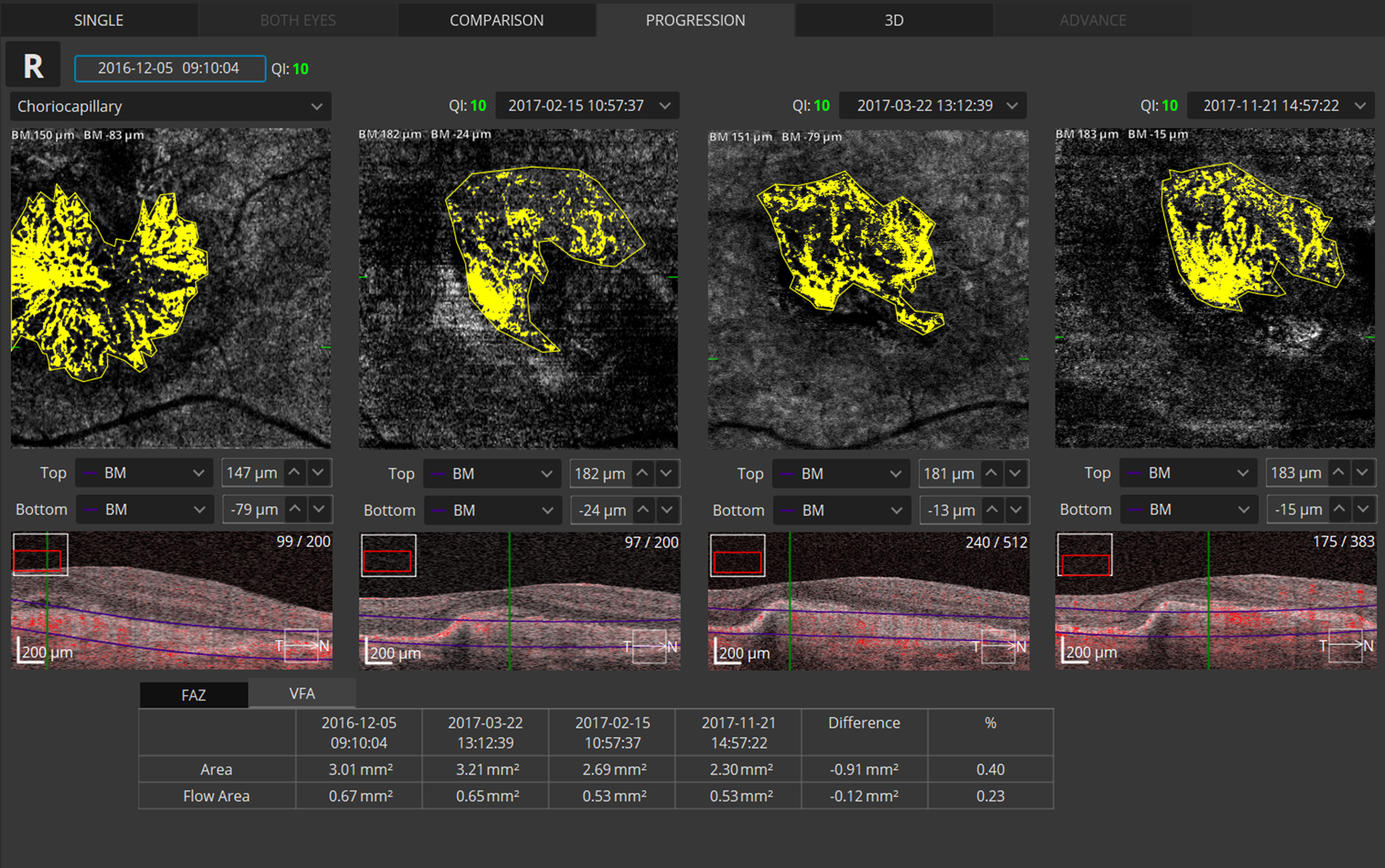
Angiography OCT
Angiography OCT provides an alternative to the traditional fluorescein method. Although OCT-A will not completely replace FA imaging, it is a quick and non-invasive tool. The software allows clinicians to observe, track, and compare changes in the microvasculature of the retina in both eyes.
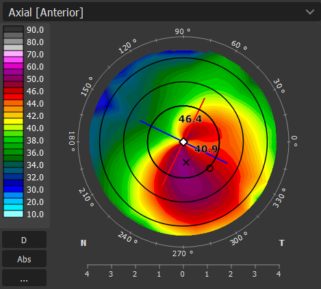
Topography
Topography OCT is a pioneering way to provide detailed corneal curvature maps. Anterior, Posterior surfaces and Corneal Thickness provide the True Net Curvature information. T-OCT™ is excellent when paired with the B-OCT® module for IOL surgery.
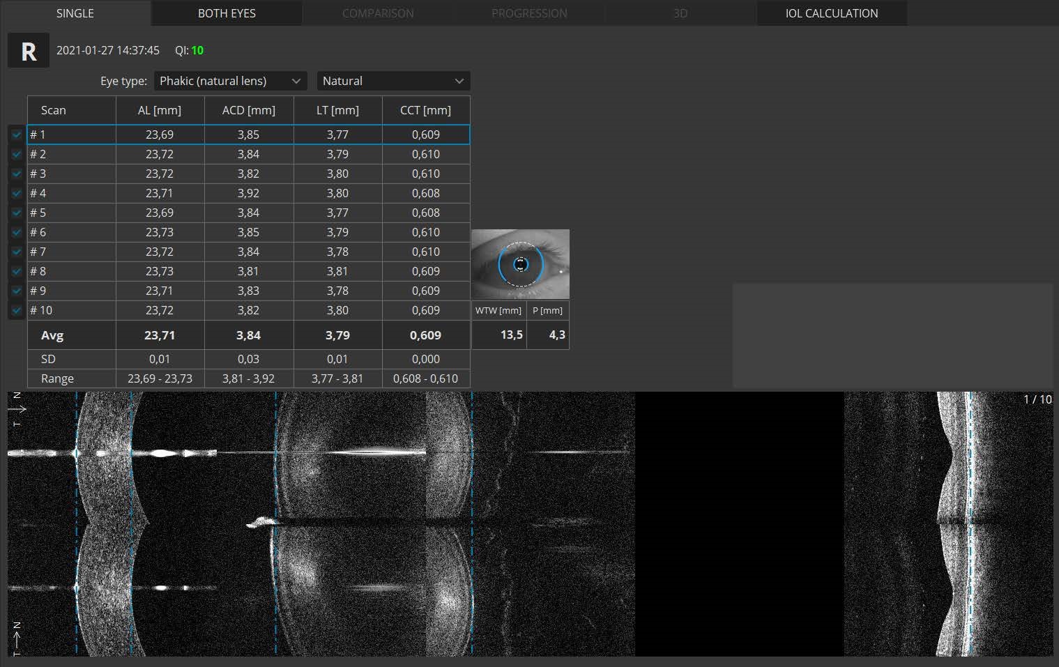
Biometry OCT
B-OCT® is an excellent tool for any clinician who manages myopia control or performs cataract surgery. Biometry OCT provides a complete set of ocular parameters: Axial Length, Central Cornea Thickness Anterior Chamber Depth, Lens Thickness, Pupil size, and White to White.
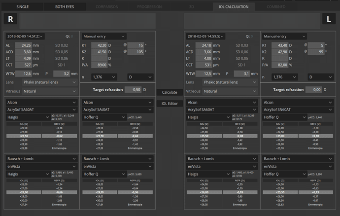
IOL Calculation
IOL formulas allow the user to calculate IOL implant parameters. The systems now support the latest IOL database standard IOLCon.org so that you can always keep your library up-to-date.
 Brochure
 Cleaning instructions
![]()
We are leaders in the industry providing you with the latest medical technology that helps grow your revenue and solidifies the relationship between you and your patients. We have four offices across Canada with local after-sales service and support for you. As well, we have an experienced, dedicated sales staff available to consult with you, ensuring all of your needs are met. We have developed relationships with suppliers from around the world to bring you reliable, high-quality ophthalmic technology, since 1980.

9820 Boulevard Du Golf
Anjou, Québec
H1J 2Y7
1 877 388-1515
111 Adam Street, Unit B
Belleville, Ontario
K8N 5K3
1 800 267-5597
#8 - 7541 Conway Avenue
Burnaby, BC
V5E 2P7
1 888 855-6558
Devices advertised may not have been licensed in accordance with Canadian law.